Wednesday, May 14, 2014

MORE ABOUT DORSOLATERAL PREFRONTAL CORTEX


My take on why manual therapy "works", part 6.

OLDER POSTS IN THIS SERIES
Part 1: IS MANUAL THERAPY EVEN NECESSARY? 
Part 2: NEUROTAGS! YOU'RE IT! 
Part 3: ABOUT "LEARNING" 
Part 4: SKIN STRETCHING AND MOVEMENT ILLUSIONS 
Part 5:  TACTILE DIRECTION DISCRIMINATION IN THE DORSOLATERAL PREFRONTAL CORTEX
NEWER POSTS IN THIS SERIES

Part 7: TREATMENT CONTEXT, NON-SPECIFIC EFFECTS  
Part 8: SOME FINAL THOUGHTS ON NON-SPECIFIC EFFECTS


..................


The next statement in the SomaSimple post is: 
6. If you already have the patient's DLPFC primed with some pain ed, you'll be able to get it to focus on skin stretch/ruffini input. 
What? How can we know that?
.........

We human primate social groomers are so accustomed to thinking bottom up that we may not even know what DLPFC even is. So this post will be a very simple tutorial about that.

What is DLPFC?
It's easy enough to look up in Wikipedia, so let's go do that. (1)

"The dorsolateral prefrontal cortex (DLPFC or DL-PFC) is an area in the prefrontal cortex of the brain of humans and primates. It is one of the most recently evolved parts of the human brain, that undergoes an extremely prolonged period of maturation that lasts until adulthood.[1] DLPFC is not an anatomical structure, but rather a functional one. This region lays in the middle frontal gyrus of humans (i.e., lateral part of Brodmann's area (BA) 9 and 46 [2] and in macaque monkeys, this region is around the principal sulcus (i.e., in Walker's area 46 [3]).[4][5] Other sources consider that DLPFC is attributed anatomically to BA 9 and 46 [6] and BA 8, 9 and 10.[1]
DLPFC is connected to the orbitofrontal cortex, and to a variety of brain areas, which include the thalamus, parts of the basal ganglia (specifically, the dorsal caudate nucleus), the hippocampus, and primary and secondary association areas of neocortex, including posterior temporal, parietal, and occipital areas.[7] Also, DLPFC is the end point for the dorsal pathway (stream) that tells the brain how to interact with the stimuli. On the other hand, the ventrolateral prefrontal cortex (located more inferior/ventral to DLPFC) is the end point of the ventral pathway (stream) that brings information about the stimuli’s characteristics.[8]
An important function of the DLPFC is the executive functions, such as working memory, cognitive flexibility, planning, inhibition, and abstract reasoning.[9] However, DLPFC is not exclusively responsible for the executive functions. All complex mental activity requires the additional cortical and subcortical circuits with which the DLPFC is connected.[10]The DLPFC is also the highest cortical area that is involved in motor planning, organization and regulation.[10]"

It's the bit we use for thinking. And socializing I guess. We human primates are usually very proud of this bit, even though we often have issues with whatever other peoples' dorsolateral prefrontal cortex comes up with or have come up with.

For example, using my own DLPFC, I can easily spot holes in other peoples' ideas about manual therapy. It's a lot harder for me to spot my own!

Anyway, this is the bit that Lundblad was viewing in the scanner when he examined his unique subject who had the iatrogenically wounded spinal cord on just one side in just one dorsal column pathway (see 
Part 5:  TACTILE DIRECTION DISCRIMINATION IN THE DORSOLATERAL PREFRONTAL CORTEX).
This is the bit he figured out had tactile direction discrimination. Yes, this area is not only cognitive-evaluative, it's also sensory-discriminative. With emphasis on discriminative.
(See Melzack's neuromatrix model (2)) 

SOURCE(3)
Here is a very nice picture of it. 
It's the purple area
right where horns would grow 
out of our heads (if we had horns). 


It does so very much much more however. It is so wired into other regions of the brain. Mostly it serves to inhibit. Everything else in there. When it wants to. Which is why we need to recruit it right up front so it can get busy busting up irrational ideation and dismantling unwanted detrimental neurotags for the long haul.

But we can also get it busy sensing what we are doing on the patient's skin surface. How cool is that!?

...................


2. Melzack R; Pain and the Neuromatrix in the Brain. Journal of Dental Education 2001,Volume 65, No. 12, 1378-1382 (full pdf)
3. Web Topic 8.7: Brains and Decision Making. Bradbury JW, Vehrencamp SL; Principles of Animal Communication 2nd Ed., online companion to the textbook Principles of Animal Communication, Second Edition by Jack W. Bradbury and Sandra L. Vehrencamp, published by Sinauer Associates.


Tuesday, May 13, 2014

TACTILE DIRECTION DISCRIMINATION IN THE DORSOLATERAL PREFRONTAL CORTEX


My take on why manual therapy "works", part 5.

OLDER POSTS IN THIS SERIES

Part 1: IS MANUAL THERAPY EVEN NECESSARY? 
Part 2: NEUROTAGS! YOU'RE IT! 
Part 3: ABOUT "LEARNING" 
Part 4: SKIN STRETCHING AND MOVEMENT ILLUSIONS
NEWER POSTS IN THIS SERIES
Part 6: MORE ABOUT DORSOLATERAL PREFRONTAL CORTEX 
Part 7: TREATMENT CONTEXT, NON-SPECIFIC EFFECTS 
Part 8: SOME FINAL THOUGHTS ON NON-SPECIFIC EFFECTS


..................


The next statement in the SomaSimple post is:

"This will engage the dorsolateral prefrontal cortex, even. (See Lundblad et al 2010, about Ruffinis and DLPFC)"

It's time to revisit that Lundblad paper (1). Actually, Olausson was involved in it, of course..
He seems to be involved in all papers that involve exteroceptive or interoceptive skinput.

Here is the full reference:

Lundblad LC, Olausson HW, Malmeström C, Wasling HB. Processing in prefrontal cortex underlies tactile direction discrimination: An fMRI study of a patient with a traumatic spinal cord lesion. Neurosci Lett. 2010 Oct 15;483(3):197-200

ABSTRACTWe have investigated cortical processing of tactile direction discrimination (TDD) in a patient with unilateral tactile disturbance due to spinal cord lesion. The patient R.A. (male, 45 years old), suffers from a traumatic dorsal column lesion at the level of Th XI-XII on the right side. He was instructed to report the direction of 2mm long skin pull stimulations applied in a proximal or distal direction on his right or left lower legs during functional magnetic resonance imaging (fMRI). Although R.A. considered himself to have nearly normal tactile sensibility, testing showed severely disturbed TDD on his right leg whereas results were within the range of healthy subjects on his left leg. For both legs TDD activated an extensive cortical network that included opercular parietal area 1 (OP1) of the second somatosensory cortex (S2), as has previously been observed in healthy subjects. However, dorsolateral prefrontal cortex (DLPFC) and anterior insular cortex (AIC) were only activated for the unaffected (left) leg where TDD was normal. A revisit of previously published data showed that healthy subjects consistently had TDD-related activations in DLPFC and AIC. However, in several healthy subjects AIC, but not DLPFC, was also activated for skin pull stimulations per se without the TDD task. Thus, the patient's data, in conjunction with the previous results from healthy subjects, suggest that DLPFC processing is important for tactile decision making based on proper tactile input.

The implications are kinda huge, actually... Here is my confirmation bias, full on, full blast:  I very much like the idea that Ruffinis (my very favourite sensory ending, way out in skin, easy to manipulate - dead easy) are somehow totally tied up with tactile direction discrimination! Not just sensing, but direction discrimination! Movement illusion!
This study demonstrated, in a single subject with an incredibly rare iatrogenic spinal cord lesion (that only affected his dorsal column pathway(3), and only on one side!..the control group was built right in!!).. that the dorsolateral prefrontal cortex activated on the appropriate (opposite) side, with skin stretch, inside an MRI machine, from skin stretch on the unaffected leg, but did not with skin stretch on the affected leg, whereas everything else in the brain activated bilaterally. Effectively isolating DLPFC as being divorced/severed from input from (specifically, one presumes) Ruffini input. By said lesion to the dorsal column pathway.

I mean, how often does an opportunity to combine all those factors into one study come along? The odds against this combination of factors plus a researcher canny enough to want to know, and willing to build a machine that would be acceptable to a scanner, plus a patient willing to go in one just for the research, after having his spinal cord injured by an epidural injection gone wrong in the first place for something totally unrelated - I mean.. sometimes the universe knocks me back on my butt, it's so marvelous at coughing up stuff like this every so often!  
It's pretty unlikely that something like this would ever be replicable. But that doesn't mean it isn't great science and great scientific deciphering.

OK, enough of the science appreciation. Please read the blogpost I linked to, where you'll see a picture of the device that the patient wore inside the scanner, and a description of the paper itself. And a lot more gee-whiz golly-gee science appreciation writing by me that you may ignore if you so choose.


1.  Lundblad LC, Olausson HW, Malmeström C, Wasling HB. Processing in prefrontal cortex underlies tactile direction discrimination: An fMRI study of a patient with a traumatic spinal cord lesion. Neurosci Lett. 2010 Oct 15;483(3):197-200


3. Overview of ascending pathways: Dermoneuromodulation: Ascending pathways Mar11/2012


Monday, May 12, 2014

SKIN STRETCHING AND MOVEMENT ILLUSIONS


My take on why manual therapy "works", part 4.

OLDER POSTS IN THIS SERIES


Part 1: IS MANUAL THERAPY EVEN NECESSARY?
Part 2: NEUROTAGS! YOU'RE IT! 
Part 3: ABOUT "LEARNING" 
NEWER POSTS IN THIS SERIES
Part 5: TACTILE DIRECTION DISCRIMINATION IN THE DORSOLATERAL PREFRONTAL CORTEX
Part 6: MORE ABOUT DORSOLATERAL PREFRONTAL CORTEX
Part 7: TREATMENT CONTEXT, NON-SPECIFIC EFFECTS 
Part 8: SOME FINAL THOUGHTS ON NON-SPECIFIC EFFECTS

The 4th statement from the SomaSimple post is this:
4. Add a novel stimulus, e.g., skin stretching (see Gandevia and Collins 2005)(1), allow the brain to enjoy a movement illusion. "Illusory movements were evoked at the interphalangeal (IP) joints of the index finger, the elbow, and the knee by stimulation of populations of cutaneous and muscle spindle receptors, both separately and together."

SKIN INPUT AFFECTS MOTOR OUTPUT

Simon Gandevia has been at this whole neurophysiology business for decades. I tried to read his papers a number of years ago (but got distracted by something else, as usual..)
Anyway, I remember one paper in which he had difficulty separating all those annoying cutaneous receptors away from all the juicy muscle spindle receptors he was actually trying to get at and measure. I think it had to do with reflex testing - you know, tapping the patellar tendon to get the quads to jump a bit. In this paper, it looks like he succeeded in isolating muscle spindles but at the same time, Collins and he decided to study cutaneous receptors as well - their effects on movement illusion.

It's a great little paper. In my life it represents a bit of a watershed paper in that it pointed out a really important thing - that the brain takes cues from receptors in skin (and this from a muscle spindle researcher!), not just from muscle. You see, back in the day, my training was completely lopsided - the nervous system was only important for, and was taught as, a motor output system. There was no emphasis on anything afferent, except from muscle spindles. Nervous system as black box connected only to muscles, input or output. Everything else, inconsequential.
Unless you are an aspiring human primate social groomer - then it would have been really nice to have learned something about how the brain codes for skin-put.

Anyway, back to the Collins and Gandevia paper. Not only does the brain take cues from receptors in skin, it creates movement illusions based only on the information it gets from skin! Remarkable. Sort of a ++ for simple human primate social grooming. Aha! So, if you want the brain to think there is more space around a certain place, like a knee joint maybe, stretch the skin there and see if the knee will bend further after. (It will, most of the time. Unless there is an actual mesodermal blockage in there, and not just a critter brain, interoceptive pain illusion that feels like a restriction.)

Gandevia and Proske published another paper (2) a few years ago about kinesthesia.
Turns out Ruffini endings contribute to proprioception. What do you know? In fact they say skin receptors are more likely to play a role in joint position sense than joint receptors do. Oh, they still love their muscle spindles, of course, but look: 

"The cutaneous receptor most likely to subserve a kinaesthetic role is the skin stretch receptor, the slowly adapting Type II receptor served by Ruffini endings (Chambers et al.1972; Edin, 1992). For kinaesthesia at the forearm, stretch of skin over the elbow during elbow flexion can provide information about both position and movement. Movement illusions generated by stretch of skin of the hand and over more proximal joints, when combined with muscle vibration were greater than when either stimulus was applied on its own (Collins et al. 2005). The authors made the point that this was not just a matter of skin input facilitating the muscle input and that cutaneous input generated by skin stretch contributed to kinaesthesia in its own right."
Now, add this together with what Olausson and his colleagues have been doing in Sweden for decades(3), and I think we have a case for skin-put having an important role in the world. Not that we're going to ever stop doing it - human primate social groomers are just doing what our own vertebrate mammal primate nervous systems have always done, since whenever they started self-organizing then interacting half a billion years ago. 


YES-CICEPTIVE INPUT

I have already blogged my fingers to the bone about Olausson and his group in Sweden. Read all about it.


  • THE FINE OLD ART OF FINGER PULLING


  • Don't be confused about this video - a lot of emphasis has been placed on the visual component of the illusion. I would argue that the kinesthetic component is equally if not more important. It wouldn't be very hard for some clever researcher type to design a test for that - blindfold one group of participants, pull their fingers in exactly the same way, in the machine. Have another group just look, don't touch them at all. Use the mirrors to make it look like their fingers lengthen all by themselves. A third group could have both illusions at the same time. 

    I bet the last group would have the best results, but hey, if the first group had pretty good results, you wouldn't need a great big expensive machine to create a movement illusion, and ordinary human primate social grooming would be vindicated.

    1. Collins D, Refshauge KM, Todd G, Gandevia SC; Cutaneous receptors contribute to kinesthesia at the index finger, elbow, and knee. J Neurophysiol. 2005 Sep;94(3): 1699-706 (full text pdf)

    2. Proske U, Gandevia SC; The kinaesthetic senses. The Journal of Physiology, 587, 4139-4146. (full text)

    3. Mind tricks may help arthritic pain

    4. Illusion could halve the pain of osteoarthritis, scientists say


    Sunday, May 11, 2014

    ABOUT "LEARNING"

    My take on why manual therapy "works", part 3.

    OLDER POSTS IN THIS SERIES
    Part 1: IS MANUAL THERAPY EVEN NECESSARY?
    Part 2: NEUROTAGS! YOU'RE IT!

    NEWER POSTS IN THIS SERIES
    Part 4: SKIN STRETCHING AND MOVEMENT ILLUSIONS
    Part 5: TACTILE DIRECTION DISCRIMINATION IN THE DORSOLATERAL PREFRONTAL CORTEX 
    Part 6: MORE ABOUT DORSOLATERAL PREFRONTAL CORTEX 
    Part 7: TREATMENT CONTEXT, NON-SPECIFIC EFFECTS 
    Part 8: SOME FINAL THOUGHTS ON NON-SPECIFIC EFFECTS
    .....................

    Moving on to point 2 and 3 in the little SomaSimple post list:


    2. Something new has to be added for the brain to get busy with. Manual contact provides it a way to distract itself from the unfortunate neurotag, long enough to get itself started on building a new representation of somatic "reality" - a new neurotag.
     3. The old one will never go away, but the brain can now "choose" to adapt itself to the new one. It's called extinction learningand happens at the receptor level of brain operation. Once that happens, old triggers are no longer triggery. 

    What do I mean by that? It's a pretty compact statement to make. There is much to unpack in it.
    Let me try.

    First off, I've convinced myself there are at least two brains in there, critter and human:
    1.  critter brain, mostly interoceptive, an internal regulation system, all the brain stuff that works from day one when we are born, even if it takes a bit longer to mature in some aspects. All the brain nuclei that keep us alive, as individuals, and later, come online to keep us alive as a species.  The part of the brain that is born knowing how to swallow, blink, burp, throw up, breathe, sleep; it's the brain that puts us to sleep at night, but keeps our heart beating and our lungs breathing. Rolls us over in the night, hopefully without waking us up. Wakes us up in the morning because it's hungry and wants us to go get it something to eat. It's the brain that runs our blood flow, our temperature, our immune system, our descending modulation to nociceptive input. It started out as a spinal cord, invented by fish, half a billion years ago. The front end of it added more hard drive as critters became more complex, faced more complex challenges, like gravity, by adapting movement behaviour to physical forces, couldn't reproduce anymore just by squirting out eggs and sperm into the water and hoping they bumped into each other. We still have all that stuff, because nature never throws out anything that works - it just adapts it to new purpose, an evolutionary principle known as exaptation.
    But be aware, the spinal cord, even though it doesn't have much hard drive, at ALL, will automatically take care of motor output business reflexively, whenever it can - that is to say, whenever it isn't being tonically inhibited by more rostral centers. Uh-oh - more words..
    "Tonically" means continually. As opposed to "phasically", which means in a punctuated manner. These words crop up all the time when you read about the brain(1).

    Tonic would be like holding your hand under a dribble of water flowing continuously from a tap. Phasic would be one drop of water falling on your hand, then another, indefinitely(2). EXAMPLE: 

    "Second, inhibition can be "phasic" or "tonic". Phasic inhibition is a short-lasting inhibition typically generated by the activation of GABAA receptors following action potentials in a presynaptic interneuron. However, there are also more long-lasting forms of inhibition. One form is the activation of GABAB receptors by spillover of GABA caused by GABA release from specialized interneurons. A second form is the "tonic" GABAA conductance activated by ambient GABA in the extracellular space (Farrant and Nusser, 2005). This form of inhibition is mediated by molecularly and functionally specialized GABAA receptors(2)."
    2. a human brain, externally driven, mostly exteroceptive, dependent on social relations and symbolic thought, the one "we" live in and consider as "I". This consists of the expansion of frontal lobes and the association cortices. The one that makes up rules to live by in consensus with other human primates, then decides that's the "right" way to live and everyone who doesn't is by definition "wrong" (or at least suspicious..)
    The human brain can be taught how to be more aware of interoception.

    Anyway, I'm not entirely sure, but pretty confident, that in the case of pain, it's the critter brain that "makes" it, or else just doesn't inhibit nociception in a timely or effective manner, then sends it forward or else simply lets the info travel all though the brain, including to the "conscious awareness" bits, where the human brain "perceives" it.

    I see my job as distracting the one while competently handling the other, so that the two might live together in harmony once more, through new learning. Each learning episode seems to take at least two minutes. I don't think any lasting change can happen in there in any shorter length of time.

    Here is where I'm just making stuff up: I can't prove any of this, all I can do is write about my thought process.
    There are many types of learning.
    Here are a group of blog posts on the topic, from Neurotonics, which developed from this one by Matthias Weinberger, The Devil is in the Details.

    I think the kind I favour for the process of manual therapy learning, kinesthetic learning, is extinction learning (3). I have no idea if it's the case, but it makes sense to me. Pain becomes tied up in fear. Fear (of moving) is involved. Fear (of moving) must be disengaged. The brain makes a new representation of its "bodymaps"every couple nanoseconds (4). 


    Stress is involved in "learning" - the right amount of glucocorticoids will facilitate learning. Too much will be harmful. "Pain" is "learned" at a synaptic level. "Pain" is reinforced through social stress, be it real or just perceived. (Which means, for us independent human primate social groomers, we can give ourselves some leeway - there are some pain mountains out there that we just aren't going to be able to climb. We can't fix some of our patients' broken lives. Furthermore, some people's pain, even though it may be biopsychosocial, may have abnormal "bio.")

    If you can provide a patient's human brain with a window of relief, in which it can move its physicality freely again, and it's a normal intact brain, it will rapidly accept/learn the new representation. Positive feedback loops are interrupted.
    The human brain can get back in charge of the critter brain. It will take back its right to move its physicality, easily. Like water rolling down a hill. Critter brain will have stopped "biting" the human brain.

    So, it's the critter brain we want to distract. How? By offering a new input. It's kind of automatic that the critter brain's salience network will come over to sniff out a new input, even innocuous. Meanwhile, we are not doing anything the human brain finds the least bit uncomfortable. Or at least we shouldn't be, in my opinion. 

    The human brain is in on the plot to contain the critter brain, because we provided it with information at the start. In this way, we can work together (psycho-social) to overcome the critter brain (bio), from top down and bottom up at the exact same time. The therapist helps from the outside by creating the treatment context, a story-line maybe, maybe about helping move/feed/drain nerves or something (that's the one I use all the time). But the patient's human brain will be doing all its own heavy lifting/changing/neuroplasticizing around the new input. 

    1. Mark Farrant, Zoltan Nusser; Variations on an inhibitory theme: phasic and tonic activation of GABAA receptors. Nature Reviews Neuroscience 6, 215-229 (March 2005)
    2. Neural inhibition. Peter Jonas, Gyorgy Buzsaki, Scholarpedia page. 
    3. Edwin Santini, Robert U. Muller, and Gregory J. Quirk; Consolidation of Extinction Learning Involves Transfer from NMDA-Independent to NMDA-Dependent Memory. The Journal of Neuroscience, 15 November 2001, 21(22): 9009-9017 4. Notes re: Interoception, from A.D. (Bud) Craig's lecture.

    Saturday, May 10, 2014

    NEUROTAGS! YOU'RE IT!


    OLDER POSTS IN THIS SERIES

    .............................................


    Earlier I posted "Is manual therapy even necessary?" Probably not, in the long run... However, it has a place in the short run. This blog series is an exploration of that.

    Yesterday I quickly scratched down thoughts with supporting evidence, and listed them in a blogpost here. The plan is to flesh out these ideas a bit better, in this blog series.

    The first point is about neurotags.
    I wrote,

    "1. The brain, once a neurotag has arisen and become easily triggered, can't go back to being a brain that never had that neurotag. (I think of a neurotag as a tangled mess of enhanced firing, sort of like a positive feedback loop but more of a birds' nest than a simple loop)"

    So, let's discuss neurotags first.

    What is a neurotag?
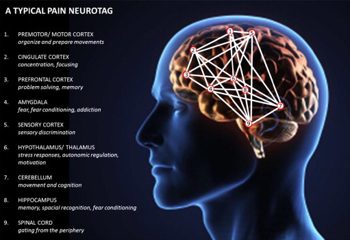
    A neurotag is a "pattern of neuron activation which creates a certain output of the brain, such as a perception, thought, movement or immune system response."(1)
    ".. when someone mentions a slipped disc, and you activate your neurotag for “slipped disc” to think about it, you also activate some of the member cells for your back pain neurotag.  Therefore thinking about a slipped disc will lower the threshold for activation of your back pain neurotag.  Get it?  Reread if necessary! This is a very simple explanation for why pain can be modified by so many different inputs."

    SOURCE: Zac Cupples (3)

    Construction of a neurotag is perfectly normal brain behaviour. It's not about your brain being defiant or evil or mean to you. It's how brains remember what they have learned - they build associations. Synapses are involved. Enhanced signalling is involved. Glia are involved, along with 250,000,000 synaptic proteins that turn over every few days (4). Imagine that.
    When I think of a pain neurotag, I think of a cobweb gathering dust. I also remember the ant swarm that got stuck in a positive feedback loop around the base of a tree, and couldn't break free (5). I think of Erik Meira's post (6) about positive feedback loops, Getting Rid of SomethingPositive.
    "Remember "positive" does not mean "good" when we talk science. It means "additive".


    .......................................

    1. Review of Moseley/Hodges Talk Part Two Todd Hargrove, BetterMovement.org blog, Oct21/2012
    2. What is a Neuromatrix? David Butler, Neuromatrix Training blog, Jan22/2008
    3. Graded Motor Imagery, Zac Cupples, April 24/2013
    4. The Remarkable Neuron: Erin Schuman at TEDxCaltech, youtube video 14:30, Feb8/2013
    5. The Ants go Marching One by One... Diane Jacobs, HumanAntiGravitySuit blog, April30/2014
    6. Getting Rid of Something Positive Erik Meira, The PT Podcast blog, April9/2014

    My take on why manual therapy "works" Part 1

    Part 1: IS MANUAL THERAPY EVEN NECESSARY?

    This series will be a longish look over several blog posts at all the ways I can think of that the nervous system of another will "feel" our own.

    Of course, manual therapy won't work (for pain) without...

    1. some sort of explanation beforehand - hopefully pain ed. that is congruent with reality and doesn't contain any tissue based nocebo.. (1) 
    2. movement therapy afterward, and suggestions re: avoiding behaviours that people unwittingly do that contribute to a pain presentation unique to them. (E.g., always standing on one leg, always sleeping on the same side, always sitting with the same elbow leaning on the same arm of the couch, always turning the head the same way to watch TV...)
    Let me be clear - manual therapy is optional. The aspects of treatment before and after manual "treatment" will work by themselves, a lot of the time. But manual therapy sandwiched in there can be optimal, in my opinion. 

    Whether it is optimal will depend entirely on how we explain what it's "for" - we need to improve the language around manual therapy!

    As Adriaan points out,
    "First, let’s discuss the assumption that pain comes from tissues. The Cartesian model that correlates tissue issues (nociception) to pain is over 350 years old, and it’s still doctrine in medicine and various therapies. The model is false. You can have tissue injury and no pain. You can have pain and no tissue injury. For too long, practitioners and patients have sought answers to their pain by exploring the various tissues, including joints, muscles, ligaments and more. Pain is leading reason that people seek care, and when they do seek help for pain, they are presented a tissue-based model to explain their pain. Think about it: A patient comes to you seeking help for pain, and you teach the patient anatomy! No wonder pain rates in the US have doubled in the last 15 years alone. Never before have we performed as much surgery or prescribed as much medicine for pain in the history of mankind, and pain rates are ever increasing. A large portion of the blame should be leveled at these outdated models. It’s time practitioners wake up and realize people in pain are interested in…pain! 
    This leads us directly to the second issue."There is also an assumption that patients are not smart enough to learn the latest neuroscience of pain. Shame on us for thinking that. Research has shown patients are in fact able to understand the biological processes of pain. Therapeutic neuroscience education (TNE) takes complex neurobiological and neurophysiological processes and explains pain to patients via metaphors, examples and pictures. We have been teaching people about pain for years, in various countries, to different age groups, in different languages, to various ethnicities, etc. The end result? They all get it. The best part is they experience less pain and disability; move and function better despite no hands-on interventions; catastrophize less; are less afraid and are able and willing to move further into pain during exercise and functional tasks. Healthcare education has simply become a display of knowledge. “Let me tell you how much I know about….” The language we use is completely foreign to patients. Even more worrisome, the current medical vocabulary contains various terms and languages that actually increase fear and anxiety. Ever been guilty of using terms like torn, ripped, instability, bleeding, rupture and so forth?"


    So, let's try to reinvent language around manual therapy.

    1. Every Chronic Pain Patient Has a Brain - Adriaan Louw


    Newer posts in this series:
    Part 2: NEUROTAGS! YOU'RE IT! 
    Part 3: ABOUT "LEARNING"
    Part 4: SKIN STRETCHING AND MOVEMENT ILLUSIONS

    Part 5: TACTILE DIRECTION DISCRIMINATION IN THE DORSOLATERAL PREFRONTAL CORTEX
    Part 6: MORE ABOUT DORSOLATERAL PREFRONTAL CORTEX
    Part 7: TREATMENT CONTEXT, NON-SPECIFIC EFFECTS 
    Part 8: SOME FINAL THOUGHTS ON NON-SPECIFIC EFFECTS